Eye Exams are for more than Vision
The eyes may be the window to the soul, but they can also tell you a lot about your health. Problems spotted in a patient’s eyes might be a signal of problems lurking somewhere else in the body. Some abnormalities might only be visible by the ophthalmologist when the eyes are dilated. Advanced Practice Nurse Dawn Specht of Shore Physicians Group in Somers Point said looking into her patient’s eyes may reveal changes that warrant further investigation.
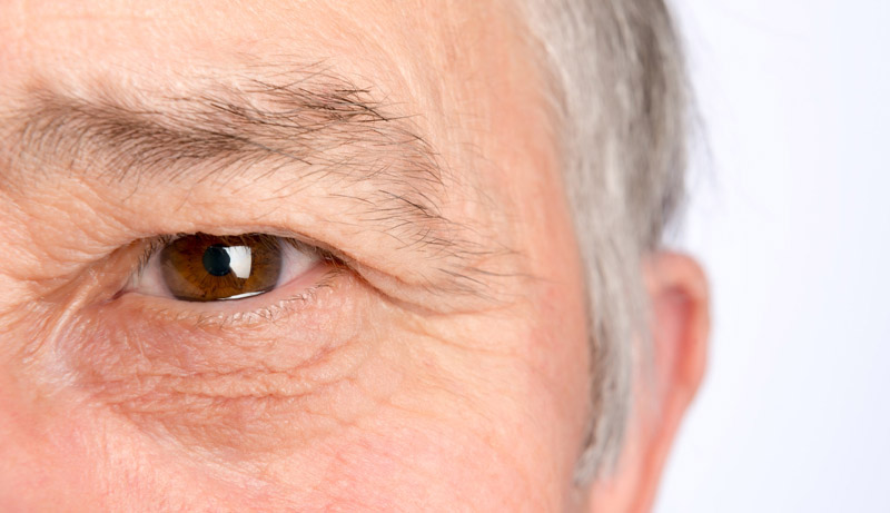
Here’s looking at you
During an annual exam, going a step beyond vital signs and evaluations and examining a patient’s eyes may go a long way to assessing your overall health. “The eye exam involves assessing exterior and interior structures. The eyes’ exterior structure is what you see when you look in the mirror,” said Dawn Specht APN. “You see your eyelids, eye lashes, sclera, or the white part of your eye, the iris, or the color portion of your eye, and the dark pupil in the center. Changes in the eye may indicate that more investigation is necessary.”
Seeing more than meets the eye
As Specht explained, one change in the eye that would cause concern is a yellowing of the sclera. “This could indicate a problem with your liver. A common problem is hepatitis or an inflammation of the liver. The inflammation could be from alcohol, infection or even your gallbladder.”
Eyelids that are red or swollen may be a common finding for an allergy sufferer. “But if you notice sediment in your eyelashes that resembles dandruff it may indicate that allergies are not the problem but eyelid inflammation, also known as blepharitis. The conjunctiva or pink lining inside your eyelid might swell and appear red, indicating conjunctivitis that could be caused by a bacteria or a virus.”
Evaluating eye movement
Your eyes have highly specialized muscles and nerves. Specht said, “The movement of the eyes is evaluated to test both the muscles and the nerves in the eye. If the pupil does not constrict and dilate smoothly or if the eyes are not able to move in all directions, further investigation is warranted. The inability of the eye to move freely might indicate pressure on the cranial nerve. That pressure could be coming from changes in the brain such as swelling, bleeding, or even a mass.”
Getting to the real challenge
The need to get to the back of the eye to examine the retina, macula and optic disc is the real challenge, according to Specht. “This is done most effectively and efficiently during a dilated eye exam with specialized equipment in the optometrist’s or ophthalmologist’s office. The back of the eye is examined to detect glaucoma, macular degeneration and retinopathy. While normal aging increases the risk of changes being detected in the back of the eye, high blood pressure and uncontrolled diabetes may also damage the back of the eye.”
During the dilated eye exam the provider may be able to detect the formation of retinal hemorrhages. “A brain bleed or a brain mass may cause swelling of the optic nerve, referred to as papilledema. Uncontrolled diabetes may cause changes in the blood vessels of the retina. They may swell, change shape or even form new branches. These changes are called diabetic retinopathy and need to be treated to prevent the permanent loss of vision,” said Specht. “The diagnosis of diabetic retinopathy tells us that we need to improve blood pressure and blood sugar control. Hypertensive retinopathy tells us we need to do a better job at controlling blood pressure. Papilledema tells us that we need to examine the brain for problems causing increased intracranial pressure.”
Diabetes and vision
Uncontrolled or undiagnosed diabetes can be detrimental to vision. Specht said, “The Centers for Disease Control identifies diabetes as the leading cause of new cases of blindness in adults. Changes in the blood vessels in the diabetic’s retina may result in swelling, fluid leakage or new blood vessel formation. These changes are referred to as diabetic retinopathy. Diabetic retinopathy has various stages and types. During the first stage there are no symptoms. This may help us understand the estimate that 1 in 29 diabetic patients over the age of forty have diabetic retinopathy. The only way to diagnose this complication and prevent vision loss is with a dilated eye exam. Over time the small blood vessels that supply the retina can be blocked by too much sugar and the eye will attempt to compensate by growing new blood vessels but the new vessels are weak and typically leak and bleed. This is called proliferative diabetic retinopathy. Treatment now exists for proliferative retinopathy but it requires diagnosis with a dilated eye exam.
To see the scope of what happens in a dilated eye exam, visit https://www.nei.nih.gov/learn-about-eye-health/healthy-vision/get-dilated-eye-exam.
Follow National Eye Institute advice
As a primary care provider, Specht said she advises patients to be wise about their eyes and follow the National Eye Institute, a branch of the National Institute of Health, and schedule a dilated eye exam every one to two years for anyone over the age of 60, or for African Americans over the age of 40 or anyone with a family history of glaucoma. For those with diabetes or high blood pressure, a dilated eye exam should be scheduled every year.
To schedule an appointment with Dawn Specht, APN, call 609-365-6200.